
INTRODUCTION

Cancer is one of the hazardous and convoluted disease. Cancer is the main source of overall demise even after a ton of headway as far as determination and treatment in the previous few years. It is anticipated that the malignancy passing rate may achieve 13.2 million by the year 2030.Cancer includes hereditary and epigenetic reconstructing of typical body cell prompting destructive cell which at last outcomes in everlasting status and uncontrolled division.
The uncontrolled cell divisions increments with increment in tumor mass bringing about metastasis and demise of entire living being because of organ failure. Metastases is one of the greatest test to medicinal administration of growth and one of the significant reason for death in malignancy patients.
Identification of the component that prompts tumorigenesis and malignancy movement helps in discovering more strong therapeutics and enhanced diagnostics. Over the previous couple of decades several growth target (proteins, compounds or receptors) has been found and therapeutics are composed in view of these targets.1The current medication accessible for disease treatment are extremely costly, profoundly inadequate, nonspecific and has number of side effects.2
Computational drug design, set up well ordered throughout the most recent couple of decades, makes it conceivable to dispose of a significant number of the previously mentioned issues. Utilizing a wide range of calculations, approximations of restricting free vitality of synthetic mixes to a sub-atomic target can be produced in silico in a quick and exceptionally shabby path, with no requirement for physical accessibility of those compounds in this progression. PC helped sedate outline in this way permits to radically accelerating the errand of growing new medications, firmly diminishes costs and empowers the quick testing of new, yet non-blended, classes of compounds. Besides, it can likewise be utilized to anticipate other substance properties of atoms, similar to their ingestion, circulation, digestion, and excretion, and along these lines accelerate tranquilize improvement by either evacuating compounds with undesired properties or by streamlining properties of found hits. Keeping in mind the end goal to make utilization of the considerable number of focal points of present day PC supported medication outline, a plenty of various calculations and readiness steps is vital, which have be to used together in enormous computational pipelines.3
One of the regions of utilization of computational drug designing is quantitative structure action (QSAR) demonstrating. A wide range of relapse and arrangement models, and additionally the info age, information administration, include choice and model approval systems under one basic structure, so the greater part of the methodology are anything but difficult to use in blend are depicted which are rapidly extensible and adaptably usable. For the field of structure-based medication outline, a quick receptor-ligand scoring capacity, a docking calculation and a three-dimensional target-particular rescoring approach was produced by the researchers. Besides, to take into account advancement of restricting free vitality gauges got by docking, another receptor-ligand rescoring strategy was executed. It utilizes the three-dimensional data (i.e., the purported postures depicting the putative ligands inside the coupling pocket) produced by docking and test restricting free vitality estimations for different mixes so as to reestablish the docking postures. In this manner, this approach, as opposed to every single other one known, considers receptor-ligand associations, their three-dimensional areas and their objective particular significance.4
The overview of entire framework is called CADDSuite (Computer-Aided Drug Design Suite). This framework contains all the algorithms and a high number of auxiliary tools, e.g. for preparation or analysis purposes. An introduction to biochemical and computational background is also described.
Drug Discovery and Development Process
Overview of Drug Design

Drug discovery and advancement is a complex, lengthy, time expending and exceptionally costly process. It includes the cooperation of different fields, for example, medicinal chemistry, pharmacology, clinical research, drug metabolism, process chemistry…..etc.
Additionally combinatorial chemistry, high throughput screening and molecular modelling assumes a fundamental part in current drug discovery process.
It takes around 7-12 years and $ 800 million to $1.8 billion to bring new lead from drug revelation to market. Initially 1,00,000 applicant compounds, hundred of preclinical animal testing and clinical trails on thousand of volunteers and patients is conveyed to recognize a solitary showcased drug. The process from the ID of new medication to the promoting is alluded as pipeline which includes following real advances.5
- Disease selection
- Target identification
- Lead identification
- Lead optimization
- Preclinical trials
- Clinical trials
Computational Background
Overview of Computer-Aided Drug Design
Computational drug designing is the advanced process of drug discovery and development of new drugs.with the advent of this computational drug designing many of the problems so associated with the rational drug designing can be overcome.This technology has helped to reduce the cost and amount of time spend to develop a new drug.

Furthermore, computer-aided drug design may also establish molecules as promising drug candidates that would never have been tested without computer-based methods, due to either the huge search-space or their initial unavailability in synthesized form. A special case of the latter reason is the in silico construction of new molecules, i.e. compounds that have not been observed in nature but were constructed on a computer manually or by an algorithm designed for this purpose. In essence, computer-aided drug design approaches try to predict properties and actions of chemical compounds by a variety of techniques, so that molecules that are unlikely to experience the desired effect on the chosen molecular target can be cast aside. Examples of such molecular properties are absorption, distribution, metabolism, excretion and toxicity (ADMET). The expected effect on the molecular target, on the other hand, is usually evaluated by a prediction of the binding free energy (or binding affinity) of the compound to the target structure. Computer-aided drug design can be divided into two major categories: ligand-based drug design and structure-based drug design.
Ligand-Based Computer-Aided Drug Design:
The ligand-based computer aided drug design (LB-CADD) approach includes the investigation of ligands known to interface with an objective of intrigue. These techniques utilize an arrangement of reference structures gathered from Compound known to interface with the objective of intrigue and examine their 2D or 3D structures. The general objective is to speak to these mixes in such a way that the physicochemical properties most important for their coveted communications are held, though superfluous data not pertinent to the communications is disposed of. It is viewed as a backhanded way to deal with medicate disclosure in that it doesn’t require learning of the structure of the objective of interest. The two crucial methodologies of LB-CADD are (1) determination of mixes in view of substance similitude to known actives utilizing some similitude measure or (2) the development of a QSAR demonstrate that predicts biologic movement from synthetic structure. The contrast between the two methodologies is that the last weights the highlights of the compound structure as indicated by their effect on the biologic movement of intrigue, while the previous does not. The strategies are connected for in silico screening for novel compound having the biologic action of intrigue, hit-to-lead and prompt medication advancement, and additionally for the enhancement of DMPK/ADMET properties.LB-CADD depends on the Similar Property, which expresses that atoms that are fundamentally comparable are probably going to have comparable properties. LB-CADD approaches rather than SB-CADD methodologies can likewise be connected when the structure of the biologic target is obscure. Furthermore, dynamic mixes recognized by ligand-based virtual high-throughput screening (LB-vHTS) strategies are regularly more powerful than those recognized in SB-vHTS
Structure based Computer-Aided Drug Design:

Structure-based computer aided drug design utilizes the three-dimensional structure of the objective of enthusiasm for request to discover aggravates that reasonable tie to its coupling pocket and could subsequently be great drug applicants. The three-dimensional structure can be acquired by either X-ray protein crystallography, atomic attractive reverberation (NMR), or by homology modelling. The last method utilizes the structure of a homologous protein that has been dictated by one of the previous techniques. Calculations for the field of structure-based medication configuration fundamentally comprise of receptor ligand docking and rescoring approaches.
The objective of receptor-ligand docking is to anticipate the binding of a ligand in the coupling pocket of a receptor, given just the 3D directions of the last mentioned and the topology (or information adaptation) of the previous. In this way, docking approaches more often than not comprise of a scoring capacity that assesses the collaboration vitality of each (moderate) posture and a calculation that creates a wide range of poses to be assessed by the scoring capacity. Scoring capacities can for the most part be isolated into learning based and observational ones. While the previous utilize a reversal of the Boltzmann factor to compute scores from the recurrence of various perceptions, the last utilize various (regularly physically inspired) terms whose coefficients are advanced utilizing a particular informational index with known binding free energies.
Chemical Classification of Anticancer Drugs
The drugs used in the treatment of cancer are classified into different classes which are described as follows


Alkylating Agents
Nitrogen mustard: eg Mustine HCl, Mechlorethamine
- Ethylene mine: eg Thiotepa
- Alkyl sulphonate: eg Busulfhan
- Nitrosourea: eg Carmustine,Lomustine
- Triazine Dacarbazine
- Platinum complex: eg cisplastin, carboplatin
Antimetabolites:
- Folate antagonist: eg Methotrexate
- Antipurine: eg 6MP,Pentostatin
- Antipurine: 5 fluorouracil,Gemcitabine


TARGETS FOR CANCER
Since convectional chemotherapy isn’t particular for malignancy cells prompting harmful reactions there is a requirement for novel specialists with high review antitumour specificity.The significant essential to grow such medications is to comprehend the objectives that these operators should attack. In late years a number of promising new anticancer medications have been produced which target intracellular pathways or extracellular cell molecules.so a portion of the major targets are recorded underneath.
Topoisomerase as anticancer target
 The expression “chemotherapy” was initially instituted by the renowned German scientist Paul Ehrlich in the mid 1900s. It was for the most part characterized as the approach of utilizing chemical to treat illnesses. In any case, these days this term as a rule alludes to treatment with the chemicals that kill the quickly dividing cells. Obviously, some non-malignancy cells likewise partitioned rather quickly, e.g., cells in bone marrow, stomach related tract, and hair follicles, and subsequently chemotherapy are for the most part considered non-particular and are called “cytotoxic.” Common chemotherapeutic specialists incorporate alkylating operators, antimetabolites, antimicrotubule agents, cytotoxic anti-infection agents, and topoisomerase inhibitors or toxic substances. A brilliant late verifiable record of the improvement of tumor chemotherapy is accessible. In this segment, we will center around topoisomerase harms since they are among the best and most generally utilized anticancer medications. For instance, doxorubicin (Adriamycin), an outstanding Top2 poison, is as yet dynamic as a first-line treatment for breast cancer, bone and soft tissue sarcomas, anaplastic thyroid tumor, bladder growth, numerous myeloma, and Hodgkin’s and non-Hodgkin’s lymphomas, and so on. Topoisomerases settle topological issues of DNA two fold helices by giving the broken strand a chance to pivot around the in place strand (named Type IB, or Top1 for people), passing one strand (named Type IA, or Top3 for people) or duplex (named Type IIA, or Top2 for people) from a similar DNA atom through the single or two fold stranded break they produced on another duplex. Up until now, there is no medication focusing on Type IA topoisomerase. All FDA-endorsed drugs focusing on Type IB topoisomerase are camptothecin subsidiaries. Topoisomerase poison act by catching the chemical as a fruitless protein DNA-tranquilize “cleavage complex” (DNA is cut in this structure), which adequately changes over the compound into a cell harm lastly prompts apoptosis of the tumor cells.

The X-beam structures of human DNA topoisomerase in complex with DNA and the camptothecin simple were resolved in 2002. The synthetic assorted variety of Top2 harms is substantially higher. There are two isoforms of Top2: Top2α and Top2β. The structure of Top2α in complex with DNA, however without tranquilize, was distributed in 2012.The structures of Top2β in complex with DNA and medications were resolved as of late. Restraint of Top2β has been perceived to be in charge of the cardiotoxicity of a few medications, e.g., doxorubicin, and along these lines has been considered as a “anti-target” for planning new topoisomerase harms.
To segregate the drug binding modes in Top2α and Top2β, we initially connected sub-atomic docking with our recently created scoring capacity to assess the coupling methods of some Top2 drugs, in particular, VP-16 (etoposide), m-AMSA and mitoxantrone. Our docking computations very much recreated the crystallographic restricting method of VP-16 of every a ternary complex of Top2β; with a root-mean-square deviation of just 0.65 Å. Sub-atomic progression reproduction of Top2β in complex with VP-16 likewise affirmed the crystallographic restricting mode. Interestingly, the compliances of Arg503 of Top2β in complex with m-AMSA and mitoxantrone from the atomic progression reproductions goes astray from their unique crystallographic adaptations, showing an unwinding procedure from the adaptations decided with the medication substitution strategy for setting up the holo protein precious stones. The coupling method of VP-16 in the cleavage complex of Top2α was controlled by the joined utilization of homology displaying, docking, and sub-atomic progression reproductions, which fell inside a comparative restricting pocket of Top2β cleavage complex. The dynamic data of Top2α and Top2β may encourage more productive planning toward Top2α-particular medications.
The binding modes in the molecular dynamics simulations are consistent with the crystallographic binding mode of this ternary complex.
Mutant RAS protein inhibitors
 In around 20– 30% of every single human tumor, transformations of proteins in the Ras (curtailed from “rodent sarcoma”) family are much of the time watched. Ras proteins are little GTPases, and are known to shape nanoclusters on films. The Nano space area and film introduction of Ras could create isoform diversity. Creating helpful operators that can return the abnormal flagging caused by the mutant Ras proteins is considered as a powerful approach for malignancy chemotherapy.
![Figure 16: The X-ray crystallographic binding mode of N-{1-[(2, 4-dichlorophenoxy) acetyl] piperidin-4-yl}-4-sulfanylbutanamide (designated as 6) bound with K-Ras G12C mutant. The hydrogen bond between 6 and Gln61 is shown as a thin yellow line](https://i0.wp.com/www.ijprs.com/wp-content/uploads/2018/09/18-1.png)
For instance, staurosporines have been found to disturb phosphatidylserine trafficking and to mis-restrict Ras proteins. Andrographolide subsidiaries have been found to hinder the GDP/GTP nucleotide trade and to revoke oncogenic Ras work. Recently, the coupling hotspots on K-Ras were related to the test based sub-atomic flow reenactments, which encourage denovo drug design of new little particles.
Kinase Inhibitor
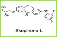

Figure 17: The X-ray crystallographic binding mode of skepinone-L bound with p38α mitogen-activated protein kinase (MAPK). The PDB accession number is 3QUE.The hydrogen bonds between skepinone and the backbone nitrogen and oxygen atoms of Gly100 are shown as thin yellow linesSupported by the 2001 FDA-endorsement of the primary kinase inhibitor, imatinib (or Gleevec), a point of reference work of disease therapeutics, kinases have turned out to be a standout amongst the most seriously sought after focuses in late pharmaceutical inquires about. Until July 2015, a sum of 28 little particle kinase inhibitors have been endorsed. In spite of the noteworthy accomplishments in the improvement of kinase inhibitors, drug resistance is as yet one of the focal issues for growing far superior therapeutics, and the unconstrained mutations in the ATP-binding space of the kinases are a standout amongst the most imperative causes.
As of late, long (18.5 μs) express solvent thermodynamics combination counts for the protection causing changes of p38α MAPK were led to accomplish great relationships amongst’s tentatively and computationally decided binding free energies.
This p38α MAP kinase was known to have an elective restricting site close to Phe169 after authoritative of diaryl urea inhibitor BIRB796, and unequivocal solvent molecular dynamics simulations could over and again recognize this mysterious binding site. A computational strategy, SCR, which unequivocally assesses receptor adaptability with the rotamer library and with full atomic points of interest, was proposed to plan kinase inhibitors. This strategy could duplicate the known binding methods of the benchmarked kinase inhibitors.
It is fairly normal that aggravates that objective kinases will tie to a group of kinases, rather than one particular kinase. Knowing the range of the kinase restricting profile is likewise vital for deducing the most reasonable utilizations of the new mixes. For this situation, not just one single protein ought to be considered as the objective of medication outline. As a rule, distinguishing proof of conceivable biomolecular focuses (off-targets) of little chemical particles is a vital advance for disentangling the hidden reasons for their activities at the sub-atomic level. To this end, we have built a web server, ID Target (http://idtarget.rcas.sinica.edu.tw) that can anticipate conceivable binding targets of a little synthetic particle by means of a divide-and-conquer docking approach, in mix with our as of late created scoring capacities that depend on vigorous relapse examination and quantum compound charge models. It was exhibited that ID Target can recognize kinase inhibitors. Scientists likewise demonstrate that ID Target can repeat known off-targets of drugs or drug like compounds, and it is conceivable that the proposed new targets could be abused for advance applications.
HDAC inhibitor


Figure 18: The binding pose of the ligand N-(4-aminobiphenyl-3-yl) benzamide (designated as LLX) in the original crystal structure of HDAC/inhibitor complex (yellow), the pose with local optimization (cyan), and the pose predicted with AutoDock4 docking with the scoring function AutoDock4RAP(pink). The root-mean-squared deviations of the two predicted ligand poses from the crystal pose are 0.315 Å and 0.277 Å, respectivelyDouble activity Compound have a place with the least complex class of “designed multiple ligands” (DMLs), which are single compounds intended to at the same time regulate numerous objectives. As a result of the engineered possibility and the medication resemblance limitations, the majority of DMLs are double activity Compounds. In the previous decade, outlining double activity Compounds has turned into a developing worldview for drug discovery. In this survey, we will center around double activity intensifies that fuse some portion of the inhibitors of histone deacetylase (HDAC) as the substance moieties.
Deacetylation of the lysine buildups of histones by HDAC brings about a profoundly minimal condition of chromatin, which makes those areas of the chromosomes have a transcriptionally inert state. HDAC overexpression has been found in an assortment of human malignancies, including myeloid neoplasia and solid tumors. The relationship of HDACs with oncogenic DNA-restricting combination proteins and other harsh interpretation factors constitutively smothers particular tumor silencer qualities. Subsequently, HDACs have been considered as a critical class of targets for disease treatment. A few HDAC inhibitors (HDACis) are as of now under clinical trials on either monotherapy or mix treatment for malignancy treatment. It isn’t astonishing to see that the idea of DMLs has been connected to plan new HDAC’s by consolidating other dynamic operators focusing on inosine monophosphate dehydrogenase, atomic vitamin D receptor, tyrosine kinase receptor or topoisomerase II 24 in growing new therapeutics for tumor medications.
A moderately less verifiable truth is that statins are likewise HDAC inhibitors. Statins, for example, lovastatin and atorvastatin, are best known to diminish serum cholesterol levels through focused restraint at 3-hydroxy-3-methylglutaryl coenzyme A reductase (HMG-CoA reductase, or HMGR). The HMGR inhibitors (HMGRis) are viably used to diminish the rate of cardiovascular and cerebrovascular issue and to forestall cardiovascular disease (CVD). Statins have a built up record of security and adequacy in human CVD counteractive action. In spite of the fact that statins have as of late been appeared to be viable for tumor avoidance in observational, preclinical, and certain randomized controlled investigations, the fundamental molecular mechanism is as yet subtle. Our examinations showed that statins specifically follow up on HDACs as inhibitors, which gives an epigenetic component to tumor aversion and malignancy treatment.
It has been accounted for that the joined utilization of anticancer specialists with statins may decrease reactions to accomplish better treatment of growths. Besides, the in vitro try utilizing a mix of HDACi and HMGRi has demonstrated some level of synergism for the enlistment of apoptosis of HeLa cells. The basic synergistic system has been proposed as takes after: the down-direction of GGTase-I β subunit, caused by HDACi (TSA in that review), upgrades the exhaustion of mevastatin-prompted geranylgeranylated RhoA.
Given the previously mentioned confirm, it is possible that simultaneous hindrance of HDAC and HMGR would be a promising methodology for growth treatment. In any case, there will be some known disadvantages if multi-component drug cocktail are adopted for example, complex pharmacokinetics, unpredictable drug to drug interactions and formulation issues because of various solvency of individual drugs.
Scientists have in this manner set out to outline a progression of double activity Compounds to target histone deacetylase (HDAC) and 3-hydroxy-3-methylglutaryl coenzyme A reductase (HMGR) correspondingly. These compounds indicated powerful inhibitory exercises against HDACs and HMGR with IC50 esteems in the Nano molar range. These compounds viably diminished the HMGR action and in addition advanced the acetylation of histone and tubulin in malignant cells, however were not dangerous to typical mouse fibroblast cells and human fibroblast cells.10
Current Status

Earlier the development of anticancer agents used to be a lengthy, time consuming and expensive process but thanks to computational drug designing which has lower the investment on technologies, time required and resources. Large amount of information available about the small molecules, target structure and advancement in genomics and proteomics has helped in application of computational drug designing every step of drug discovery and development .The 3D structure of target and chemical compounds have increased the affinity towards the target with the aid of computational methods. The protein structure so determined by X-ray crystallography and NMR technique can be used as drug target and help to design target inhibitors which can fit in the binding pocket of the protein. some of the examples of successful application of computational drug designing are listed below.11

Tubulin is a protein which polymerised to form microtubule which is a component of eukaryotic cytoskeleton. It plays an important role in cell proliferation and division and also a major target in anticancer therapy. Tubulin inhibitors are developed by ligand based drug designing in order to prevent the cancer cell division.
Prior the improvement of anticancer agents used to be an extensive, tedious and costly process however on account of computational drug designing which has bring down the venture on innovations, time required and resources. Large measure of data accessible about the small molecules, target structure and progression in genomics and proteomics has helped in use of computational drug designing in each progression of drug discovery and advancement .The 3D structure of target and synthetic compounds have expanded the proclivity towards the objective with the guide of computational methods. The protein structure so dictated by X-RAY crystallography and NMR strategy can be utilized as medication target and help to configuration target inhibitors which can fit in the coupling pocket of the protein. some of the cases of fruitful use of computational medication planning are recorded below.
Tubulin is a protein which polymerised to shape microtubule which is a segment of eukaryotic cytoskeleton. It assumes an imperative part in cell multiplication and division and furthermore a noteworthy focus in anticancer therapy. Tubulin inhibitors are produced by ligand based drug designing so as to stop the cancer cell division.
P53 unregulated modulator of apoptosis (PUMA) is an apoptotic protein having a place with the group of Bcl-2 protein which is managed by tumor silencer p53 and causes apoptosis. PUMA inhibitors are created by structure based pharmacophore modelling keeping in mind the end goal to represses apoptosis.
Table 1: selected inhibitors developed with computational designing
| Compond Name | Therapeutic Area | Function | Approvals |
| Gefitinib | NSCLC | EGFR kinase inhibitor | 2003 |
| Erlotinib | NSCLC
pancreatic cancer |
EGFR kinase inhibitor | 2005 |
| Sorafenib | Renal cancer
Liver cancer Thyroid cancer |
VEGFR kinase inhibitor | 2005 |
| Lapatinib | ERBB2- positive breast cancer | EGFR inhibitor | 2007 |
| Abiraterone | Metastatic castration -resistant prostrate cancer or hormone refractory prostrate cancer | Androgen synthesis inhibitor | 2011 |
| Crizotinib | NSCLC | ALK inhibitor | 2011 |
Computational drug designing has generated large amount of new anticancer drugs and are a milestone in drug discovery.12
FUTURE PERSPECTIVES
With the expansion in the accumulation of data about the little atoms and biomolecular structure utilizing structure and ligand based approaches, there is an improvement in computational medication planning of anticancer drugs. In future better comprehension about etiology of infections would help in the revelation of new medication targets and furthermore blend of targets. The whole natural framework can’t be recreated on the computer so endeavors ought to be made to include all the conceivable parameters. Unavailability of reliable experimental information and confinements in toxicity expectation display are a portion of the difficulties looking by computational drug designing. Advanced toxicity forecast model ought to be produced so as to evaluate the poisonous quality of drug in kidney, liver, lung, heart and other organs. Mechanism of diseases, genomics and proteomics, new drug targets, natural leads, physiochemical properties and so on should be recognized later on so as to get huge accomplishment in growth treatment.13
CONCLUSION
Computational drug designing techniques have extraordinary potential in drug discovery especially in lead distinguishing proof and lead optimization. It is an advanced, convenient and quickened strategy for sedate revelation and development. Numerous of therapeutic agents have been computationally intended to treat cancer, so computational drug discovery and development holds a great promise for future growth of anticancer drugs.
With the regularly expanding accumulation of biomolecular structures, constant upgrade of computational power, and enhanced correctnesses in displaying the sub-atomic communications at the nuclear level, it is expected that calculation will play a significantly more vital part in the drug discovery process sooner rather than later. Better understandings of the etiology of illnesses with the assistance of biological system and frameworks pharmacology additionally prompt distinguishing proof of new medication targets and furthermore imaginative combination of drug targets for designing new drugs all the more viably.
REFERENCES
- Carels, N., Tilli, T., & Tuszynski, J. A. (2015). A computational strategy to select optimized protein targets for drug development toward the control of cancer diseases. PloS one, 10(1), e0115054. https://doi.org/10.1371/journal.pone.0115054
- Ferreira AK, Kawamura B, Jorge SD, de Azevedo RA, Zaim MH, et al. (2017) Developing Novel Anticancer Drug Candidates Regarding the Integration of Three Main Knowledge Fields: Computer-Aided Drug Design, Chemical Synthesis, and Pharmacological Evaluation. J Drug Des Res, 4(2): 1035.
- Sugrue, M. F. (2000). Pharmacological and ocular hypotensive properties of topical carbonic anhydrase inhibitors. Progress in retinal and eye research, 19(1), 87-112. https://doi.org/10.1016/S1350-9462(99)00006-3
- Monroe, D. (2007). Looking for chinks in the armor of bacterial biofilms. PLoS biology, 5(11), e307. https://doi.org/10.1371/journal.pbio.0050307 , PMid:18001153, PMCid:PMC2071939
- Gao, C. (2016). Computer-aided drug design approaches in developing anti-cancer inhibitors.
- Sliwoski, G., Kothiwale, S., Meiler, J., & Lowe, E. W. (2014). Computationalmethods in drug discovery. Pharmacological reviews, 66(1), 334-395. https://doi.org/10.1124/pr.112.007336 , PMid:24381236, PMCid:PMC3880464
- Muegge, I., & Martin, Y. C. (1999). A general and fast scoring function for protein− ligand interactions: a simplified potential approach. Journal of medicinal chemistry, 42(5), 791-804. https://doi.org/10.1021/jm980536j, PMid:10072678
- Böhm, H. J. (1994). The development of a simple empirical scoring function to estimate the binding constant for a protein-ligand complex of known three-dimensional structure. Journal of computer-aided molecular design, 8(3), 243-256. https://doi.org/10.1007/BF00126743, PMid:7964925
- Acharya, C., Coop, A., E Polli, J., & D MacKerell, A. (2011). Recent advances in ligand-based drug design: relevance and utility of the conformationally sampled pharmacophore approach. Current computer-aided drug design, 7(1), 10-22. https://doi.org/10.2174/157340911793743547 , PMid:20807187 , PMCid:PMC2975775
- Lin, J. H. (2016). Review structure‐and dynamics‐based computational design of anticancer drugs. Biopolymers, 105(1), 2-9. https://doi.org/10.1002/bip.22744, PMid:26385494
- Prada-Gracia, D., Huerta-Yépez, S., & Moreno-Vargas, L. M. (2016). Application of computational methods for anticancer drug discovery, design, and optimization. Boletín Médico Del Hospital Infantil de México (English Edition), 73(6), 411-423. https://doi.org/10.1016/j.bmhime.2017.11.040, https://doi.org/10.1016/j.bmhimx.2016.10.006, PMid:29421286
- Dev Bukhsh Singh (2014) Success, Limitation and Future of Computer Aided Drug Designing. Transl Med (Sunnyvale) 4: E-127. doi:10.4172/2161-1025.1000e127
- Durrant, J. D., & McCammon, J. A. (2011). Molecular dynamics simulations and drug discovery. BMC biology, 9(1), 71. https://doi.org/10.1186/1741-7007-9-71 , PMid:22035460, PMCid:PMC3203851